"The Old Achilles"
How many of us have returned after a break from running and the old Achilles has reared it's ugly head??
It's all too common for distance runners and increasingly so with changes in volume, intensity, surface type and biomechanics (especially for those embarking on the barefoot trend!) to experience Achilles pain.
Let's have a look at what commonly occurs physiologically to the Achilles when they start to degenerate and the treatment options available.
Basic Anatomy
The Achilles tendon, as the name suggests is a strong tendon composed of type 1 collagen converging from the gastrocnemius superiorly and the soleus (calf muscles) to form a strong attachment to the posterior calcaneus (heel bone). The tendon itself spirals around to it's attachment almost turning a full 90 degrees during it's course. It does this so that when it has to withstand tensile load it is able to 'unwind' allowing the collagen fibre to eccentrically lengthen and withstand large forces. Tendons withstand tensile stress extremely well however they do not absorb compression or shear forces nearly as well.
In a healthy Achilles tendon type 1 collagen predominates with small amounts of the thicker (yet less resistant) type 3 collagen. The anatomy under the microscope would look white in colour and have a uniform arrangement with type 1 collagen arranged in tight linear bundles.
When the tendon starts to degenerate due to a variety of factors (more on this later) the tendon undergoes pathologic changes, some of the changes that might be seen are;
- Thickening of the Achilles region
- Palpable nodules (particularly the mid portion)
- An influx of neo-vessels (colour doppler ultrasound)
- Collagen disorganisation (ultrasound)
- Thickened areas of type 3 collagen infiltrating into the tendon substance
- Swelling and crepitus
- Tenderness to palpation in the mid portion and in some cases the insertion to the calcaneus
- Mild to moderate atrophy in the gastrocnemius and soleus
- There are also usually musculoskeletal imbalances further up the chain
Some of these changes would be quite obvious and some tendons may exhibit changes with no symptoms. For the most part and from a clinical stand point most symptomatic tendons do show some of these changes along with a history that would suggest an overload type pattern of training. These changes are often coupled with the more mature athlete (40 years +). As we age our tendons change slowly in microstructure to a greater predominance of type 3 collagen which as we saw earlier become more disorganised in their alignment and with stand less tensile stress.
So what are the causes?
One of the key points to understand about Achilles tendinopathy is that rarely is this a condition of inflammation. The title of achilles tendinitis is a bit of a misnomer when you consider that most cases of Achilles tendinopathy do not show signs of inflammatory markers. Tendinitis does occur however, it is not as common as once thought and usually responds well to appropriate treatment.
We still do not fully understand all the causes of Achilles tendinopathy however there are some factors relating to runners that are fairly consistent
- Pronation (rate/speed of pronation)
- Biomechanics - fore foot landing (Achilles load!)
- Increase in volume
- Increase in intensity
- Poor recovery time (this usually co-incides with the above 2 points)
- Poor dorsiflexion (less than 5 cm)
- Poor end range stiffness (lack of myofascial load bearing ability)
- Over-striding - this increases the load on the Achilles
These factors combined with an ageing tendon may cause changes to occur over time resulting in pain and altered gait.
So how can this be treated?
One thing we do not fully understand about Achilles tendons is why some recover quite quickly and others with the same findings and similar treatment do poorly. There are various forms of interventions and many will depend on what type of presentation and how long the Achilles has been problematic. An acute bout of Achilles pain if caught early usually responds quite well with load modification, heel raises (5mm), soft tissue therapy, joint mobilisation and addressing any pelvic or gait imbalances.
More chronic cases will usually need more time and modification of the running program and more intensive treatment. If we think about the causative factor in the development of the problem then we will go along way to addressing the case and modifying those factors
In recent years, Hakan Alfredson popularised the advent of eccentric calf training after research he conducted in 1998 with subsequent papers showing promise with short and long term results. This was further supported by world leaders in tendon research such as Prof Jill Cook and Prof Nicola Maffulli.
Alfredson's program was as follows:
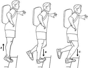
Three sets of 15 reps of the straight leg heel drops and three sets of 15 reps of the bent leg heel drops, at a speed of 4 sec up and 4 sec down twice per day for 12 weeks and to continue despite mild pain (usually a subjective level of about 5/10 on a VAS scale). Once the pain starts to subside the load is increased usually about 2-3kg every 5-7 days. Once the load becomes easier again the load is increased. The key to eccentric loading is moderate controlled progressive overload, that is the load must be controlled and consistently increased to cause a microstructure adaptation in the tendon.
Alfredson's original research had chronic Achilles subjects loading with up to 55kg on their back!! Clinically it can be found that once the load increase to an appreciable amount (15-20kg) results start to show good results clinically. A duration of 8-12 weeks is usually warranted to see results, in the case of minimal results over this time and in the absence of other progressive changes then further investigation may be warranted.
This has been a mainstay of conservative therapy over the last decade. Dr Jill Cook further advised heel drops not below parallel for the de-conditioned athlete ie. from the floor level. Eccentric heel drops appear to be most effective for Achilles pain in the mid portion, that is, 2-6cm above the insertion onto the calcaneus (heel bone). Eccentric training is less effective in cases of insertional Achilles pain largely due to the fact that we are dealing with a multitude of tissues (bursa, fat pad, type 2 collagen, boney tissue) hence degeneration of this area is a lot more complex.
So what changes have been found in the degenerate tendon after a period of successful eccentric training?
Alfredson studied the microstructure of tendons before and after 12 weeks of eccentric training and found the pain free tendons had a more organised collagen arrangement (= better tensile load ability) and a decrease in neo-vscularisation (nerve and blood vessel influx into the tendon substance).
These heel drops also increase muscle bulk to the calf muscle which is often atrophied on the symptomatic side. In some cases, clinically the Tibialis anterior becomes over active decreasing the capacity of the opposing posterior calf muscles to activate overloading the Achilles tendon. A course of soft tissue therapy to the over active side can help the posterior calf muscles to increase their activation, hence reducing load on the Achilles.
In the case of a thickened Achilles soft tissue mobilisation can be an effective adjunct. The aim of such therapy is to mobilise the thickened fibres in response to maladaptive loads. It is thought that mobilising the fibres increases fibroplasia thus, creating increased turnover of type 1 collagen and again creating a more robust tendon.
Further treatment involves looking at the role pelvic position and stability play in the role of foot position and pronation. Poor pelvic biomechanics will place an increased load on the load bearing structures of the lower limb.
There are numerous conservative treatment approaches beyond the scope of this article however the following may be a piece of the rehabilitation model
- Polidocanol (nerve sclerosant)
- Autologus blood injections
- Platelet rich plasma injections (PRP)
- GTN patches
- Cortisone injections (very controversial)
- Orthoses
- Kinesio taping
There is a plethora of information available in journals, sports medicine texts and well reference books. This is a small compilation from the clinical observations and experience in dealing with Achilles tendinopathy that I have had over the years

A. Straight leg heel raise
B. The eccentric portion
C. Bent leg eccentric - this is often forgotten in many eccentric loading programs
Be sure to complete both straight and bent leg exercises. The straight places a bias on the upper calf portion whilst the bent leg exercise places a bias on the lower calf portion


 |
| Figure 2 |
The area above is typical of mid portion achilles tendinopathy (2-6cm above insertion)

Figure 3
The area above (figure 3) is typical of insertional tendinopathy however in reality this involves a more complex tissue structure (bursa, fat pad, tendon, connective tissue). Hakan Alfredson of Sweden and world renouned Achilles researcher reported a lower success rate with insertional tendinopathy

Figure 4
Figure 4 (above) shows marked blood flow changes on a color doppler representung neovascular bundles often implicated in chronic tendinopathy in achilles tendons. Applying a nerve sclerosant via ultrasound guided injection often abolishes the nerves and the pain in most cases (Alfredson 2006).
No comments:
Post a Comment