Tuesday, 31 July 2012

Hamstring Tendinopathy: A case of long-term management

A Case Study


A 30 year old female with a history of hamstring tendinopathy spoke to me at length yesterday about her hamstring tendinopathy (I have been a part of the process along the course of treatment) and explained her treatment and investigation over a course of 17 months (and still continuing).

In April (2011) during a standard 20km long run our subject feels pain in her left hamstring that is quite localised, however it does not stop her completing her long run. Later that week the hamstring pain progresses to a point where she could not run.

Initially she rests for a week and attempts another run only to have the pain return more severely and to a point where she must walk home. A further month is taken off from running and no other treatment is sought. It becomes obvious to her this is longer term and treatment will be needed. She is unable to run and wakes with morning pain and stiffness.

In June of last year she saw a soft tissue therapist who found her left glute med wasn’t functioning properly (poor timing), her TFL was overloaded and her iliopsoas (hip flexors) were also doing a lot of work. She was unable to activate her hamstring on a ‘bridge’ * assessment. Treatment to the above areas improved her bridge and glute timing and pain, but only temporarily.

Fig 1. The hamstring functions eccentrically on and just before heel strike. Large loads are place through the hamstring complex. If the hamstring experiences pain and/or dysfunction load will be place through the tendon and not taken up by the muscle itself

A moderate, controlled eccentric program was trialled for the hamstring, increasing weight weekly and tolerating pain. After 8 weeks there was no improvement.

In August of last year, a referral to a sports doctor found, through ultrasound some thinning of her tendon (though this was bilateral – could this just be ‘her anatomy?’ Does this predispose her?). Cortisone was injected into the tendon without guidance. There was no change in her pain and she was still unable to run.

In November (3 months after the first cortisone injection) she paid a second visit to the sports doctor where an MRI was performed on her lumbar spine for possible referral. This reveals a perfect spine for a healthy 31 yr old. She is recommended chiropractic intensive treatment, consisting of twice per week treatment for 8 weeks. On cessation her back felt great however her hamstring pain had no change.

A second opinion was sought in March of this year (2012) where a referral for another ultrasound was made, finding extensive degeneration about the hamstring tendon and thickening between the tendon and the surrounding paratendon. An ultrasound-guided cortisone injection with saline was injected into the paratendon space in an effort to ‘debride’ the surrounding tissue.

Fig 2. Tendinopathy usually occurs in the attachment of the long head biceps femoris. The junction between the muscle and tendon is also a weak spot (MTJ) leading to degenerative tears. 


At the same time an appointment was made to see a physiotherapist with experience in this area. He initially provided low-level glute strengthening and hydrotherapy.

There is a plan to start some progressive overload eccentric training as we speak. Eighteen months down the track this is still long and ongoing and with no certainty with outcome.

There are numerous clinical cases in the distance runner where hamstring tendinopathy is long and drawn out. This is one injury that needs to be addressed as soon it appears. As tendon research and understanding improve over the years, so should our clinical outcomes.

For now,

Jimmy

jimmybarker9@gmail.com

Saturday, 28 July 2012

Tibial Nerve as a Source of Plantar Heel Pain


Plantar heel pain is a common presentation to the healthcare practitioner dealing with musculoskeletal pain. Distance runners incur a high incidence of lower limb injuries. Falling into this category is plantar heel pain. A lengthy discussion has already been put forward in a recent blog, however it is important to consider other causative factors involved with the onset and treatment of plantar heel pain. This article will consider the tibial nerve (it’s branches) as a source of pain in the heel often mimicking plantar fascia-itis (fascia-oses)

The Tibial nerve actually makes a portion of the infamous sciatic nerve in the posterior thigh (along with the common peroneal nerve). It becomes superficial in the posterior knee before going deep as it descends to supply most of the calf musculature. The nerve then becomes superficial again, medial to the ankle before branching as it exits the tarsal tunnel*

The nerve we are most concerned with clinically is the medial calcaneal branch in close proximity to the attachment of the plantar fascia on the inside of the calcaneus (heel).  If compressed, tensioned or irritated it can be a source of pain presenting as plantar fascia-itis. 

Due to it’s proximity to the calcaneus it is often considered that the attachment point for the plantar fascia, (what runners might discuss as ‘heel spurs’) is the culprit. Clinically, the medial calcaneal nerve can be seen to be playing a role as the source of pain.

Fig 1. Location of medial calcaneal nerve 


So how do we differentiate between nerve involvement and the plantar fascia?

Assessment in conjunction with symptoms should guide the thought process. On assessment you will usually find reproduction of pain/symptoms, the same location or a difference from one side to the other. Symptoms may include pain at the plantar fascia attachment (calcaneus), pain extending across to the lateral side of the heel, pain in the medial ankle (tarsal tunnel). Taking the nerves through their range of motion and performing a tensioning type movement will usually elicit a response that is close to ‘their pain’. Pain reproduction is a clinical pearl when it comes to musculoskeletal assessment.

Further, there is less involvement with the connective tissue as a source of pain often resulting in less morning stiffness than would be expected if plantar fascia were directly involved. There may also be neural symptoms further up the chain behind the knee and into the glutes, eluding to neural pathology. This kind of pathology may also warm up during the course of a session

What might be occurring?

Nervous tissue likes to move and slide within the surrounding tissues (known as the mechanical interface). When nerves become compressed or ‘tensioned’ they generally receive a decreased blood flow and/or become inflamed causing pain.

Altered biomechanics, old injuries (scar tissue) and areas of increased muscle tone are common culprits. Treatment needs to focus on factors ‘driving’ the neural irritation (causative factors) whilst helping to calm the down the nervous system.

Posture as a Factor

Posture is commonly talked about but often not addressed. Efficient running posture is essential in allowing the nervous system to transmit impulses and slide within the adjacent tissues. For example if a distance runner has a seated work place job usually placing increased ‘tension’ on the nervous system as they slump down and then assumes a similar (kyphotic) position whilst running an enormous load will be placed on the nervous system.

Efficient running posture is less costly from an injury point of view. Because the nervous system is continuous, the site of pain may not always be the focus of treatment. If we go back to our seated runner we can consider limited mobility (kyphotic) through the thoracic spine to place increased tension and demands on the lower limbs an. therefore the medial calcaneal nerve!

Fig 3. mobilising the tibial and sciatic tract may give rise to increased medial calcaneal mobility. Angling the foot towards the outside will further stretch the target area


Further to this a tight nervous system will play a significant role especially in activities that are repetitive in nature!

Treatment

Treatment needs to address the nervous system as a whole - this may include;
  • Running posture
  • Seated postural options (for those in a poor seated position)
  • Neural tightness
  • Pronation (rate and amount)
  • Pelvic stability (as a factor in foot control)
  • Programming factors – recovery, volume, intensity 

Fig 2. Kyphosis of the thoracic spine from seated to running posture places a large stress (tension) on the thoracic spine


Do nerve conduction studies play a role?

In subclinical states and acute nerve pathologies, nerve conduction studies are generally not indicated (or useful) as they do not detect nervous system dysfunction at this level. They may be useful for failure of a nerve to activate or chronic pathologies where encroachment of a nerve will lead to total dysfunction of an area.

Further investigation

In chronic cases, where previous soft tissue injury around the ankle/foot has been a problem this may be a valuable adjunct. Ultrasound is able to detect if neural tissue is compressing on certain neural structures, in this case the medial calcaneal nerve. As previously discussed nerves like to slide and move through the adjacent tissue

·       The tarsal tunnel is a tunnel formed by the underlying bone (calcaneus) and the retinaculum (connective tissue). Structure’s traversing through this area are vulnerable to compression and reduced mobility

For a full list of neural mobilisation visit the NOI website www.NOI.com
There are also numerous sites on Youtube and social media devoted to this (sift through the not so good sites!!)

Please contact me if you require further information on jimmybarker9@gmail.com

Monday, 23 July 2012

A Case Study


Clinical Perspectives on Nerve Pain in Runners

Today I saw a 16 year old male elite runner who sustained a calf injury 8 days ago. He complained of pain and tightness about his left lateral posterior calf that had stopped him running for a few days.  For the most part it felt ‘tight’ but seemed to warm up.

The difference between a nerve based injury and a small tear (strain) may not always be obvious. Some of the consequences of soft tissue injury have been dealt with in a previous post Consequences of soft tissue injury. The following, details the assessment and treatment approach that should allow him to resume full load training within 1-2 weeks, depending on how well he responds to treatment.

On assessment the following was found (we’ll keep it simple);

Dysfunction of the SIJ on the left,
Increased tone in the left TFL (usually as a result of decreased glute medius stability),
Increased tone on the left gluteus maximus (again trying to take over the role of glute medius),
Tight through left Psoas (hip flexors),
Pain reproduction on a nerve tension test assessing the common peroneal nerve,
Further palpation revealed pain directly of the common peroneal nerve to the fibula head.

A thorough musculoskeletal assessment was able to define nerve pain from a calf strain despite similar presentations.  For this runner, the causative factor lay in his tight SIJ/pelvic mechanics on the left side.  Why this happened would need a look at his biomechanics when running.

Nerve pain generally warms up reasonably well (unless it’s really irritated) where as calf strains do not and simply get worse. Nerve based pain in the calf usually doesn’t evoke pain on active or passive contraction and stretch where as a tear can certainly present this way

The treatment addressed his pelvic mechanics whilst dealing with the symptoms (nerve related pain). Treating the site of pain would simply not address the problem. Home-based treatment relies on daily nerve mobilisation, decreasing the overactivity of gluteus maximus and TFL through self massage techniques. Addressing these factors is able to keep the SIJ mobile.


                                      You can see the common peroneal nerve wrapping around the head of the fibula

I’d like to hear your opinion or questions

For now,
Jimmy

Friday, 20 July 2012

Plantar Fascia Pain

This is one of the most common clinical presentations when it comes to distance runners. These cases are often long and drawn out, with patients often bouncing from one health care professional to the next.  One of the most vital pieces of information I can give to the patient is that of education on the possible causes, management and treatment. Education allows the patient to become active in the rehabilitation protocol.

Brief Anatomy

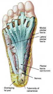
The plantar fascia is a thick band of connective tissue with investing layers from the calcaneal (heel) tuberosity to the base of the metatarsals, creating a mobile yet stable soft tissue foundation for the medial and transverse arches of the foot.

The plantar fascia is composed of type 1 collagen  (a thick protein arranged in parallel bundles) with the ability to resist high loads of tensile stress. It is considered a non-contractile tissue responding to the mechanical loads placed upon it. It has poor ability to stretch much beyond 4% of its resting length. The plantar fascia is prone to degenerative changes in response to chronic overuse and increasing with age related changes in the connective tissues.

There are 3 nerves that travel through the various layers of the plantar fascia from the medial (inside) portion of the ankle. The medial calcaneal nerve, the lateral plantar nerve and the medial plantar nerve all arise from the larger tibial nerve. These often play a role in plantar fascia pain with the medial calcaneal nerve often commonly involved as it wraps around the tuberosity. 

Anatomy of the plantar fascia

Function

The plantar fascia maintains the longitudinal and transverse arches of the foot and also serves to dampen pronation. As the foot contacts the ground and continues through to mid stance the plantar fascia lengthens under load almost causing a breaking effect, thus absorbing load. This sets the stage for the foot to then supinate creating a more rigid structure for toe off.

If the plantar fascia undergoes degenerative changes (collagen disarray, neovessel in growth, fibrosis etc) the structure become less mobile and less resistant to tensile load. This creates further overload on already compromised structure.

Onset

Plantar fascia pain usually appears with a gradual onset. Initially there is stiffness (and pain) on arising only to warm up 5 minutes later. The pain gradually progresses to pain on warm up and sometimes warm down. There is often a period of weeks to months where the patient does not seem to regress. Often this is where the running athlete does not present for treatment thinking “it’s not getting worse!”. Despite no notable regression the connective tissue may still degenerate under repetitive loads.

Over time if the athlete continues to run there will be a point where pain is constant through out the run. This is usually the first time a clinician will see the runner often months into the process.

Causes

The causes are likely multifactorial:
  • Increase in volume
  • Increase in intensity
  • Change in training surface
  • Change in footwear (or lack of)
  • Reduced dorsiflexion
  • Excessive pronation
  • Reduced great toe extension

Pronation – a possible causative factor


Prognosis

The prognosis for plantar fascia pain is less reliable than for a straight - forward muscle tear. The quicker the injury is addressed the better the outcome. Plantar fascia pain is similar to a tendinopathy in nature, with degeneration of the connective tissue a classical finding under ultrasound. Depending on the location within the plantar fascia, will depend on the type of tissue you are dealing with. For example, a degenerative enthesopathy (insertion onto the tuberosity of the heel) will involve various connective tissues, possibly fat pad and underlying soft tissues. If the focus is in the body of the plantar fascia it may be a case of the connective tissue composition (type 1 collagen).

If addressed early, clinical evidence would suggest you may be looking at 6-12 weeks recovery.

Chronic ongoing plantar heel pain injuries can be as long as 6 months to 2 years with high re-occurrence rates.

“The Doc says I have heel spurs!”

A basic understanding of the anatomy helps to understand the pathogenesis. As stated, the plantar fascia attaches to the calcaneal (heel) tuberosity. In some cases abnormal mechanics may place an increased demand upon the attachment leading to an increase in bone loading, hence, size of the tuberosity.

There is no evidence to suggest that an increase in bone (the so-called heel spurs) is a causative factor in pain. In fact some asymptomatic patients (no symptoms) have enlarged calcaneal tuberosities. It is therefore important to look to the causative factors and ways of managing plantar fascia heel pain. A ‘spur’ is not a sign of plantar fascia pain. The painful site is usually close to the attachment and may be involved in some cases.

Treatment

Plantar fascia pain presents in a similar fashion to a tendinopathy and as such should be treated as one. There is little evidence to suggest inflammation to be a factor in plantar fascia heel pain. In this case the treatment protocol will be lengthy. Addressing the causative factor(s) is paramount.

Treatment options include:

Soft tissue therapy to focal areas of thickening especially in the posterior lower limb if reduced dorsiflexion is a factor.  Joint mobilisation and local stretching may improve the outcome. Clinically local soft tissue work to mobilise thickened areas within the plantar fascia may be warranted.

Controlling excessive pronation with strengthening of tibialis posterior (inside of calf) and gluteus medius (lateral hip) or with the use of orthoses. Biomechanics during the gait cycle may have an important role to play. Overall reducing load on the plantar fascia. Low dye taping may help in the short term as a means of gaining proprioception and further reducing load

Further soft tissue treatment may help with overloaded structures (areas of increased tone) that alter gait as a compensatory mechanism or as a cause of the injury itself.

Address footwear* (or lack of – think barefoot running) - this is a hot topic at the moment! Whichever way you decide to go, make it a slow transition and be sure to use progressive over load and recovery techniques in your training program.

Consider consequences further up the chain eg; reduced torso rotation can create compensation through increase rotation of the femur, tibia and pronation. The net effect is loading of the plantar fascia. Consider the whole kinetic chain.

Low dye taping may help

What about rolling on a golf ball?

This may work to minimise the symptoms temporarily, however it will do little to help with the driving factors. Very rarely do we find in clinical practice the plantar fascia to be tight. It is important to consider the anatomy and function when considering the goal of any treatment. The plantar fascia is a thick piece of connective tissue with very little stretch – do we really need to try and stretch it?

Is it always plantar fascia involvement?

This discussion has focused on pain in the plantar fascia. There are numerous causes of pain in the plantar surface of the foot, the location will be a key to further possibilities such as:
  • Fat pad contusion (heel)
  • Plantar fascia tear (grade?)
  • Sesamoiditis
  • Morton’s neuroma
  • Stress fracture of the cuboid
  • Stress fracture of the calcaneus
  • Medial calcaneal nerve compression (common)

Summary

Plantar fascia pain is common in runners and often lengthy to treat. By the time the running athlete has presented to healthcare professionals there is usually a long history. Plantar fascia pain is degenerative in nature thus occurring over time. An earlier treatment program will likely give a better prognosis.

·       If you would like to read some great articles on the science for and against bare foot running go to the science of sport website www.sportsscientists.com

For those interested in further reading Clinical Sports Medicine, Brukner and Khan, 2007, have some excellent information and practical tips

For now,
Jimmy

Monday, 16 July 2012

Calf Strain


Calf strain was touched on in my one of my previous posts on consequences of soft tissue injury, however because it such a common injury for runners it’s worth a slightly more in-depth look.

Quite basically the calf muscle has two parts to it: the upper portion consists of the gastrocnemius (these are the power muscles you see in sprinters) and the soleus which is the endurance part of the calf which attaches to the achilles tendon.

The most common site of an acute calf strain is the medial head of the gastrocnemius (upper portion) or the junction between the upper and lower parts ‘the gastro-soleul junction’. They may also occur in the junction between the tendon and muscle (musculotendinous junction).

Onset

Calf strains usually appear during the course of one or two runs and make it impossible to run. These do not warm up and simply get worse. If you have a strain that does warm up it is more than likely to be neural tightness/irritation.

Causes
  •         Increase in intensity (faster interval training)
  •            Increase in volume
  •            Change of training surface (track/road)
  •            Decrease in recovery time (fatigue short and long term)
  •             Poor dorsiflexion (calf range of motion)
  •             Previous injury (calf)
  •             Pronation (amount and rate)
  •             Biomechanics
  •             Neural tightness (are you  ‘wound up’ through the neural structures 

Treatment

As I will always say, you need to find the causative factor involved in the calf strain – calves rarely ‘just go’ for no reason especially in such a repetitive load based sport such as running. There may be more than one factor.

Soft tissue therapy can be applied to focal areas of thickening and increased tone. Gentle joint mobilisation techniques and stretching can be applied to areas of restricted range of motion.  Neural mobilisation and self-guided nerve stretching techniques are often helpful in the recovery from a calf strain to reduce potential fibrosis around a nerve (tethering)

Biomechanics – control of hip/knee/foot is paramount to optimal stability and decreased injury risk. The gait cycle is quite complex and it may be a good idea to have a biomechanical analysis if you have access to this as part of your management. Remember, running is a skill - some pick this up well while others do not.

Just because someone has been running for along time it does not mean they have acquired the skill – you can become efficient at being dysfunctional!

Management

Management relies on identifying the factors that cause the calf problems in the first place.  Let’s look at the possible causes and pair them up with the appropriate treatment options:

  • Increase in intensity  manage appropriate recovery with programming
  • Increase in volume  manage appropriate recovery with programming
  • Change of training surface  monitor surface type
  • Decrease in recovery time  monitor load and employ recovery techniques such as hot/cold therapy and water therapy
  • Poor calf range of motion  soft tissue therapy, joint mobilisation, stretching   
  • Previous injury  soft tissue therapy to areas of focal thickening, maintain neural mobility. Strengthening the area of previous injury will also help with a strong mobile scar
  • Pronation  glute medius and tibialis posterior strengthening, orthoses (podiatrist)     Neural tightness  self guided neural stretching
  • Biomechanics  professional advice/musculoskeletal screening 
Site of strain

Important factors in the prognosis and recovery time are the site of the tear, the size of the tear and the type of tear (longitudinal, oblique, horizontal). Let’s briefly look at these:

The site of the tear is important due to the types of tissues involved and their recovery times. A tear in the musculotendinous junction will involve tendinous and muscle tissue (as the name suggests) recovery time is increased.

The size of the tear is quite obvious - more tissue separation needs a longer healing time as there is more tissue destruction to deal with. The response from an inflammatory point of view will be larger.

The type of tear is extremely important. For example we have seen in our clinic a 15 cm tear in the soleus that was longitudinal in direction with symptoms of mild pain. Conversely a small 3 cm tear in a horizontal direction across different tissue can be extremely painful with a lengthy recovery time. A similar case is likely with a tear in an oblique direction.

The treatment and management of acute calf strains lies in those factors that cause or contribute to the injury.

Until next time,
Jimmy

Saturday, 14 July 2012

Medial Tibial Traction Periostitis

"I’ve got shin splints doc”! Of all the terms that stick with injuries, this is one of the die hards.  The term ‘Shin splints’ is very broad and does not aptly describe the underlying process. A more appropriate term is medial tibial traction periostitis (Brukner & Khan 2007).

For ease of discussion it will be referred to as medial tibial pain.

Medial tibial pain is a common presentation to health professionals on a daily basis and usually arrives as a chronic case. The typical distance running scenario is ongoing for 6 months or more with many homecare interventions such as rest, stretching of the calves, anti-inflammatory medication, heat application, new shoes and the cycle continues.

Anatomy

The lower limb interacts and is largely affected by the pelvis above (biomechanics are huge in this) and affects the positioning of the foot. The 3 major tissues that play a role in medial tibial pain in the lower limb are the soleus (lower calf) attaching onto the posterior portion of the tibia and fibula, the flexor digitorum longus (these flex the toes) also attaching to the posterior tibia and the tibialis posterior attaching to the posterior tibia and fibula and attaching the navicular  (above medial arch) and base of metatarsal (base of toes)

These muscles attach to the bone via an attachment onto the periosteum. The periosteum is susceptible to large friction and traction forces in the lower one third of the tibia.

Onset
The more chronic case may be more resistant with a higher re occurrence rate. A chronic case may be 3-6 months with some cases lasting for up to 2 years

Treat this early - your options are;
ext-align: justify; text-justify: inter-ideograph;">
With the continuation of training, pain will gradually increase to the whole training session.  Ultimately it can stop training altogether.

Causes

The usual loading factors play a large role in the onset of medial tibial pain such as;


  •       Increase in volume
  •       Increase in intensity
  •       Fatigue (usually involved with the above factors)
  •       Increase in hard surface running
  •       Excessive pronation
  •       Biomechanics – stride length, pelvic position, pelvic stability
  •       Decreased recovery time between training bouts

What is very important to understand is the periosteum covers the bone as an attachment point for muscles and other connective tissues.  Continual loading and tension on this site can lead to bone strain – a pre cursor to bone stress reactions. Early intervention is the key.

Treatment

From a clinical perspective the most important aspect is knowing the causative factor(s), though in the chronic case it is likely to be multifactorial.

Knowing the anatomy and the traction forces being placed on the periosteum of the bone we can start to understand what might be occurring. Tibial bending and torsion are large in the distal tibia during locomotion. During the gait cycle uncontrolled pronation during mid stance places an eccentric ‘pulling’ on the periosteal junction. This continual ‘pulling’ causes an inflammatory response evoking painful stimuli.

Addressing the eccentric forces and the control (rate) of pronation is often helpful. This can be achieved by strengthening of the tibialis posterior and flexor digitorum longus, thus decreasing the traction forces on the tibia.

Pronation control is also aided by adequate pelvic stability during the gait cycle. Further, pronation is affected by overstriding which in turn may be reliant on pelvic angle. It is always prudent to address biomechanics when medial tibial pain is experienced.

Soft tissue therapy may be helpful in areas of focal thickening and increased tone. Joint and soft tissue health can also be assessed as a possible factor.

Prognosis

In a fresh case (less than a few weeks) and the appropriate management usually there is a good response in approx. 2-4 weeks

The more chronic case may be more resistant with a higher re occurrence rate. A chronic case may be 3-6 months with some cases lasting for up to 2 years

For the best prognosis catch it early!!

Is further investigation warranted?

If you have been on the treatment merry-go-round and your pain is not improving (or worsening) then it might be wise to visit a sports physician who may be able to order some imaging studies.  A range of imaging studies may be helpful.

Bone scan can be handy in detecting possible stress reactions however, according to Dr Tom Cross (Sydney based sports physician) one bone scan is equivalent to a radiation dose of 69 chest X-rays. That’s a big dose in one sitting! MRI’s may be effective in detecting local oedema and any soft tissue changes.

Even with further investigation, return to running is based on clinical features rather than return to normal appearance on imaging (imaging features may take months to years to return to normal despite absence of signs and symptoms) - it’s also expensive!

In summary

Treat this early - your options are;

  •       Ice
  •       Short term anti-inflams may be helpful
  •       Load/program modification (rest may be helpful initially)
  •       Biomechanics
  •       Taping and foam application (de-loading tibial area)
  •       Soft tissue therapy
  •       Tibialis posterior strengthening
  •       Pronation control
  •       Stretching exercise, if it is identified there are restricted soft tissues
  •       Joint mobilisation


Figure 1 - The tibialis posterior becomes superficial in the medial (inside) ankle. You can see how weakness in this will predispose to pronation causing a 'traction' effect on the medial tibial border. This often irritates the periosteum.


Figure 2 - The tibialis posterior tendon can be seen in the lower one third of the tibia on it's course to the attachment under the foot.

Strengthening


Figure 3 - Ideally strengthening would be performed in a standing position. Keeping the heel and the ball of foot on the ground lift the arch up 1-2cm (this will be hard to activate at first), then slowly lower. To increase difficulty hold this position and lift the heel 2-3cm off the ground, then slowly lower.

If you have a rigid foot in a static position you will probably have less available movement. These are stabilsing muscles therefore they will probably do better with multiple stes and reps throughout the day ie; 4 sets 4-6 reps (building to 10+) 3-4 times per day.


Figure 4 - Another option is to use a small step as shown aboveand slowly lower the inside of your foot off the step and then slowly raise it back up so the sole of the foot will be facing in towards the opposite leg.

All in all, an injury that you definitely want to address early rather than push through

Please let me know your experiences with ‘shin splints’ and what worked for you!